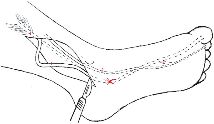
Care is taken to obtain good exposure of these two tendons most notably of the two distal fdb slips inserted on the lateral aspects of the middle third of p2. The diseased ptt is typically cleaned up or removed to eliminate it as a source of pain.

Flexor digitorum longus fdl tendon transfer to posterior tibial tendon.
fdl tendon. The fdl tendon perforating and fdb tendon perforated are identified. A flexor digitorum longus tendon transfer is indicated for patients with dysfunction of the posterior tibial tendon where the tendon has become stretched beyond its functional length or has ruptured. This muscle serves to curl the second third fourth and fifth toes.
At its origin it is thin and pointed but it gradually increases in size as it descends. The fdl tendon was pulled through the bone tunnel from plantar to dorsal and tensioned. The flexor digitorum longus tendon is transferred into the inferomedial aspect of the navicular bone and fixed in place using the tenodesis screw.
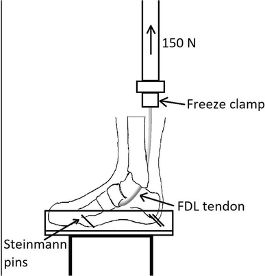
The fdl is one of the tendons responsible for bending the toes down to the floor. Edited by matthew buchanan md. All tendons measured 4 or 45 mm.
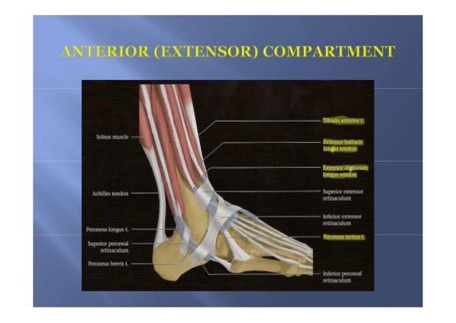
In doubtful cases traction is applied to the deep tendon to check. We found significant improvement in vas score sf 36 physical component summary pcs and lower extremity functional scale lefs. The flexor digitorum longus is situated on the tibial side of the leg.
A bicortical bone tunnel was made from plantar to dorsal ensuring the tunnel diameter matched the fdl tendon diameter. In this surgery the fdl tendon is moved from its usual position and transferred to the navicular bone. This is a flexor digitorum longus tendon transfer to the posterior tibial tendon for a dysfunctional posterior tibial tendon.
As a result the fdl tendon is transferred into a bone tunnel in the navicular bone and fixated with a screw from the bio tenodesis screw system. This video is in 4 parts. This helps support or replace the diseased ptt in order to improve function.
As the tendon passes forward in the sole of the foot it is situated above and crosses from the lateral to the medial side of the tendon of the flexor digitorum longus to which it is connected by a fibrous slip. The pulley system is opened over the mtp joint to expose the fdb superficial and fdl deep. Flexor digitorum longus fdl tendon transfer represents a surgical option in the treatment of chronic achilles tendon disorders.
This procedure is indicated for patients with a dysfunction of the posterior tibial tendon when the tendon is either stretched out beyond its functional length or when the tendon has ruptured.

 Mri Anatomy Of Ankle
 Anatomical Study Of The Neurovascular In Flexor Hallucis Longus
 Five Comprehensive Solutions For Tendon And Ligament

 Flexor Digitorum Longus Tendon Transfer To The Navicular Tendon To
